Thoughts on Science
the blog of Danelle Haake
the blog of Danelle Haake
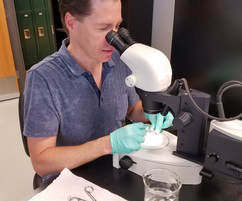 Jason working at the microscope. Jason working at the microscope. A few weeks ago, Dr. Jason Knouft, the PI of the lab where I work, was busily setting up a microscope and a variety of sterilized tools. Most of the readers of this post will not appreciate how unusual this is. While I'm spending much of my time these days at a microscope to identify the many aquatic invertebrates I've collected, Jason's research efforts are generally spent either collecting fish in rivers or analyzing data using GIS (for my non-tech/science readers, we'll say fancy mapping). While many wise graduate students would run and hide when such unusual activities begin in the lab, I elected to observe quietly to see where the day would lead. Soon the lab bench held not only the microscope and tools, but also a container of live fish and a cooler with liquid nitrogen. At this point, I knew I would be witnessing an interesting event. Jason was going to be collecting fish tissue for some baseline genomic work. Soon, Dr. Warren from Washington University arrived and the tissue collection began. I grabbed my phone and started taking pictures to document the process. The fish were anesthetized before being sacrificed to expand our knowledge of fish genetics. Next, brain tissue was removed, packaged, and frozen to preserve the DNA. Since these little fish have small brains, extra muscle tissue was collected and frozen, just in case. All in all, the dissection and tissue collection from 3 fish only took about 20 minutes. The work to read the DNA of these fish will take quite a bit longer. 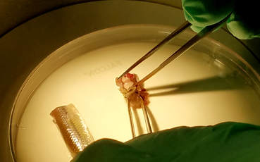 Dissection of a fish head; the white tissue being removed is the brain of a bluntnose minnow.
0 Comments
Leave a Reply. |
AuthorDanelle Haake, Archives
March 2020
Categories
All
|
 RSS Feed
